Description
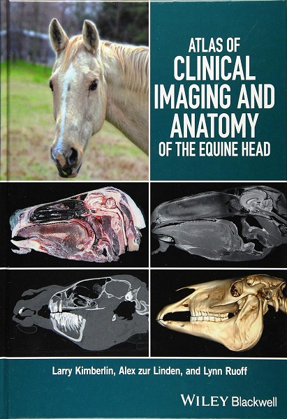
Atlas of Clinical Imaging and Anatomy of the Equine Head presents a clear and complete view of the complex anatomy of the equine head using cross-sectional imaging. Provides a comprehensive comparative atlas to structures of the equine head. Pairs gross anatomy with radiographs, CT, and MRI images, presents an image-based reference for understanding anatomy and pathology. Covers radiography, computed tomography, and magnetic resonance imaging technologies. 160 p.

- Larry Wayne Kimberlin. DVM, FAVD, DAVDC, CVPP. His extensive experience includes treatment of pets, horses, and livestock; he is also certified to perform TPLO (Tibial Plateau Leveling Osteotomy) surgery to repair cruciate ligament ruptures in dogs. ‘Crossroads Veterinary Clinic’, Greenville, TX (USA).
- Alex zur Linden. DVM, DACVR. Associate Professor in the Department of Clinical Studies, University of Guelph, Ontario Veterinary College, Guelph, ON (Canada); and Cofounder of 3DVETMED, University of Guelph, Ontario Veterinary College: a consortium for 3D printing in veterinary medicine.
- Lynn Ruoff. DVM. ‘Kittrell Animal Hospital’, Kittrell, NC (USA).
- Publication date (digital version): 2016-09 – Wiley-Blackwell; Copyright © 2017 by John Wiley & Sons, Inc.








You must be logged in to submit a review.