Welcome to vet library

Home > BASICS > Anatomy-Physiology > Atlas of Clinical Imaging and Anatomy of the Equine Head

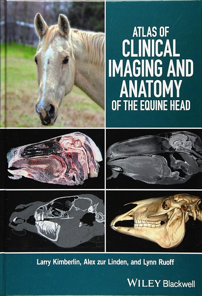
Atlas of Clinical Imaging and Anatomy of the Equine Head

45,00 

Atlas of Clinical Imaging and Anatomy of the Equine Head presents a clear and complete view of the complex anatomy of the equine head using cross-sectional imaging. Provides a comprehensive comparative atlas to structures of the equine head. Pairs gross anatomy with radiographs, CT, and MRI images, presents an image-based reference for understanding anatomy and pathology. Covers radiography, computed tomography, and magnetic resonance imaging technologies…

3 downloads

Description

Atlas of Clinical Imaging and Anatomy of the Equine Head presents a clear and complete view of the complex anatomy of the equine head using cross-sectional imaging. Provides a comprehensive comparative atlas to structures of the equine head. Pairs gross anatomy with radiographs, CT, and MRI images, presents an image-based reference for understanding anatomy and pathology. Covers radiography, computed tomography, and magnetic resonance imaging technologies. This ebook compares the gross anatomy, bone window CT, soft tissue window CT, and MRI of multiple transverse, sagittal, and dorsal sections of the head. Everything, even the smallest structures, is labeled and color coded on each view, so you know exactly what you’re looking at. The ebook also discusses how CT and MRI work and which is best for imaging certain tissues or structures. 160 p.

PREVIEW

Authors expertises affiliations

  • Larry Wayne Kimberlin. DVM, FAVD, DAVDC, CVPP. His extensive experience includes treatment of pets, horses, and livestock; he is also certified to perform TPLO (Tibial Plateau Leveling Osteotomy) surgery to repair cruciate ligament ruptures in dogs. ‘Crossroads Veterinary Clinic’, Greenville, TX (USA).
  • Alex zur Linden. DVM, DACVR. Associate Professor in the Department of Clinical Studies, University of Guelph, Ontario Veterinary College, Guelph, ON (Canada); and Cofounder of 3DVETMED, University of Guelph, Ontario Veterinary College: a consortium for 3D printing in veterinary medicine.
  • Lynn Ruoff. DVM. ‘Kittrell Animal Hospital’, Kittrell, NC (USA).
  • Publication date (digital version): 2016-09 – Wiley-Blackwell; Copyright © 2017 by John Wiley & Sons, Inc.
Additional information

Type

2 reviews for Atlas of Clinical Imaging and Anatomy of the Equine Head

5.0 out of 5 stars (based on 2 reviews)
Excellent100%
Very good0%
Average0%
Poor0%
Terrible0%


9 September 2024

This ebook is very interesting and thorough. I would consider it a must have for equine practitioners or anyone interested in equine anatomy. Although there are many books discussing the anatomy of equine viscera or limbs, there are not many that discuss the anatomy of the equine head, at least not in great detail, and there none that show the various types of imaging of the head.

Avatar for Kristal L Gebhard
Kristal L Gebhard
15 November 2017

In addition to the anatomic atlas, the book contains a well-written introduction that explains the basic principles of CT and MRI interpretation and considerations for image acquisition. Readers unfamiliar with CT and MRI will gain an understanding of what influences tissue appearance in images acquired by the use of those modalities, and appropriate references are provided for readers who wish to learn more. This book will be useful for radiologists, surgeons, internists, and other practitioners who need to acquire diagnostic images of the heads of horses

Avatar for Derek Cissell
Derek Cissell