Welcome to vet library

Home > ANIMALS > SMALL ANIMAL-EXOTIC PET > Pathology > Clinical Atlas of Small Animal Cytology

Clinical Atlas of Small Animal Cytology

45,00 

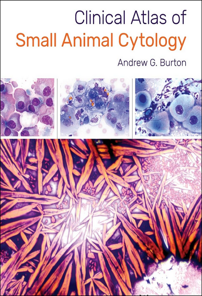
Clinical Atlas of Small Animal Cytology provides an essential guide for interpreting cytologic samples to diagnose small animal patients. Provides multiple images to differentiate cells from lesions that could look similar; features photographs of diseases with a diagnosis confirmed by pathognomonic cytologic features, histopathology, special stains, microbial culture, or other confirmatory tests. This resource presents more than 500 representative high-quality images, and emphasizes characteristic features of each disease and distinguishing features…

Description

Clinical Atlas of Small Animal Cytology provides an essential guide for interpreting cytologic samples to diagnose small animal patients. Provides multiple images to differentiate cells from lesions that could look similar; features photographs of diseases with a diagnosis confirmed by pathognomonic cytologic features, histopathology, special stains, microbial culture, or other confirmatory tests. This resource presents more than 500 representative high-quality images, and emphasizes characteristic features of each disease and distinguishing features. Key features include: – photographs of diseases with a diagnosis confirmed by pathognomonic cytologic features, histopathology, special stains, microbial culture, or other confirmatory tests; – emphasizes characteristic features of each disease and distinguishing features; – provides characteristic features of each disease and distinguishing features; – and presents more than 500 representative high-quality images. 365 p.

PREVIEW

Authors expertises affiliations

  • Andrew G. Burton. BVSc, DACVP, Clinical pathologist. IDEXX Laboratories in North Grafton, MA (USA).
  • Publication dates (digital version): 2017-08.
Additional information

Type

1 review for Clinical Atlas of Small Animal Cytology

5.0 out of 5 stars (based on 1 review)
Excellent100%
Very good0%
Average0%
Poor0%
Terrible0%


25 August 2024

I just bought this book through my specialty veterinary college site. I am a veterinary clinical pathologist and very familiar with the other small animal (dog/cat) cytology textbooks out there. I am very pleased to add this book to my library and find it a refreshing layout compared to the other textbooks. The best features include its simple and easy to find organization and outline format with salient features of each entity listed in bullets, rather than having to read paragraphs of discussion to find the key points. The cytology pictures are excellent quality, large size, and show not just one picture of a given entity but often two variations of the same thing, which is helpful. I know this book will be a microscope side resource for me, other pathologists (boarded and in-training), as well as practicing veterinarians/technicians looking at their patient cytology samples.

Avatar for M. Wiseman
M. Wiseman