Welcome to vet library

Home > BASICS > Anatomy-Physiology > Color Atlas of Veterinary Anatomy: The Horse, 2nd Edition

Color Atlas of Veterinary Anatomy: The Horse, 2nd Edition

30,00 

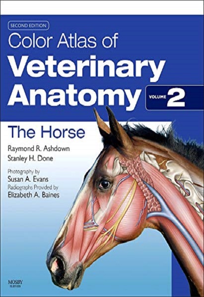
The Color Atlas of Veterinary Anatomy volume 2 presents a unique photographic record of dissections showing the topographical anatomy of the horse. With this ebook you will be able to see the position and relationships of the bones, muscles, nerves, blood vessels and viscera that go to make up each region of the body and each organ system. This volume in digital version is packed with full-color photographs and drawings of dissections prepared specifically for these texts…

2 downloads

Description

The Color Atlas of Veterinary Anatomy volume 2 presents a unique photographic record of dissections showing the topographical anatomy of the horse. With this ebook you will be able to see the position and relationships of the bones, muscles, nerves, blood vessels and viscera that go to make up each region of the body and each organ system. This volume in digital version is packed with full-color photographs and drawings of dissections prepared specifically for these texts. Key features include :Accessibly and systematically structured with each chapter devoted to a specific body region; – Important features of regional and topographical anatomy presented using full color photos of detailed dissections; – dissections presented in the standing position; – presents anatomy in a clinical context. This ebook is a must for everybody who needs to know about topographical anatomy, helpful for any practitioner who wants to know more. The Illustrations of surface anatomy are extremely helpful, as well. 362 p.

PREVIEW

Authors expertises affiliations

  • Raymond R. Ashdown. BVSc, PhD, MRCVS. Emeritus Reader in Veterinary Anatomy. University of London (UK).
  • Stanley H. Done. BA, BVetMed, PhD, DECPHM, DECVP, FRCVS, FRCPath, Visiting Professor of Veterinary Pathology. University of Glasgow Veterinary School (UK); Former Lecturer in Veterinary Anatomy, Royal Veterinary College, London (UK).
  • Publication date (digital version): 2011-06 – Mosby (imprint of Elsevier); Copyright © 2011 by Elsevier Ltd.
Additional information

Type

2 reviews for Color Atlas of Veterinary Anatomy: The Horse, 2nd Edition

5.0 out of 5 stars (based on 2 reviews)
Excellent100%
Very good0%
Average0%
Poor0%
Terrible0%


7 April 2019

This book is a fantastic learning guide and very correct reference for horse anatomy. The text is easy to understand for a lay person but still professional terminology used. Has a great index to help find what you need. The most important aspect I found is that the pictures were labelled clearly in great detail so that you can easily see what you would like to look at

Avatar for Michelle Allen
Michelle Allen
7 April 2019

I am a first year vet student at a Top Tier program here in the US. I have found this book extremely helpful for my Large Animal Anatomy course. The full color photographs are easy to understand. I love how on each page there is a full color photograph of a REAL horse cadaver along with a drawn outline of the photos that is labeled. Not only does it make it super easy to understand exactly what each structure you are look at is, it also is great for quizzing yourself because you can cover up the diagram and just look at the photograph and see if you can still name the structures.

Avatar for Marilyn Connor
Marilyn Connor