Welcome to vet library

Home > PATHOBIOLOGY > Pathology > Color Atlas of Veterinary Pathology, 2nd Edition

Color Atlas of Veterinary Pathology, 2nd Edition

35,00 

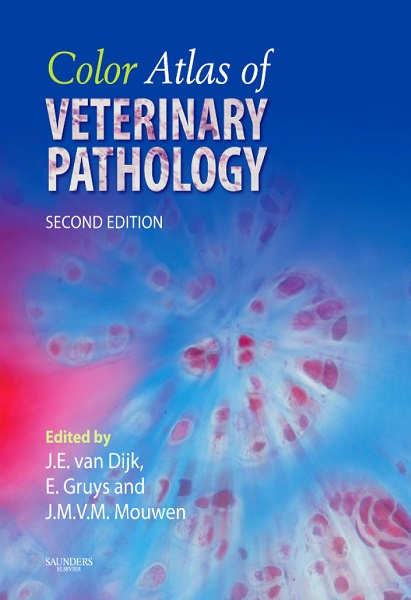
For over 20 years, the first edition of Color Atlas of Veterinary Pathology provided veterinary students and pathologists with an invaluable fast and structured survey of the complete field of veterinary pathology. Now in its second edition, the authors have thoroughly revised, updated and added to both images and text, with the focus still on domestic animals. Each chapter now begins with a short, descriptive text on each body system covered in the atlas…

Description

For over 20 years, the first edition of Color Atlas of Veterinary Pathology provided veterinary students and pathologists with an invaluable fast and structured survey of the complete field of veterinary pathology. Now in its second edition, the authors have thoroughly revised, updated and added to both images and text, with the focus still on domestic animals. Each chapter now begins with a short, descriptive text on each body system covered in the atlas. It supports understanding of disease and disease processes by visualizing how cellular pathology, inflammation, circular disturbance and neoplasia are expressed in the different organs and tissues. For this purpose it demonstrates the general morphological reactions of organs and tissues using examples from specific veterinary pathology. Key features include:Unique and internationally recognized color atlas in veterinary pathology; – encompasses all species of domestic animals; – and  a comparative approach which provides better understanding of the general mechanisms operating in the different organs. 192 p.

PREVIEW

Authors expertises affiliations

  • J.E. van Dijk, Editor. DVM, PhD in Veterinary Science. Professor and Head of Department of Pathobiology, Utrecht University, Faculty of Veterinary Medicine, Utrecht (The Netherlands).  
  • Erik. Gruys, Editor. DVM, Dipl ECVP, PhD.
  • J.M.V.M. Mouwen, Editor. DVM, Dipl ECVP, PhD.
  • Publication date (reprint original edition 2006 to digital version): 2017-12.
Additional information

Type

6 reviews for Color Atlas of Veterinary Pathology, 2nd Edition

5.0 out of 5 stars (based on 6 reviews)
Excellent100%
Very good0%
Average0%
Poor0%
Terrible0%


22 April 2018

An amazing book with great colorful pictures. It begins from how to prepare histologic sections to the description of the most important tissues, organs and systems of eight species (dog, cat, horse, cow, sheep, goat, pig, and chicken). Before the pictures, the book gives you a very useful and brief chapter introduction. It also has a detailed glossary containing nearly 750 words. A necesary book for a veterinary student. El mejor libro de histologia veterinaria que he visto!

Avatar for Andres Sanchez
Andres Sanchez
22 April 2018

A complete version of animal histology including all species!!! I love the quality of the book, the great quality of all photomicrographs!!!

Avatar for Liliana
Liliana
22 April 2018

In its succinctness, this atlas has value. There is no extraneous fluff to detract from the main points; concise text and gorgeous histological photos.

Avatar for Colorado DVM
Colorado DVM
22 April 2018

Photos are in color, which is fantastic and hard to find. Many species. Great book for your reference shelf or to help you through a class.

Avatar for Candy Matthews
Candy Matthews
22 April 2018

Very helpful for histology lab. Great colored pictures. I like the brief summaries at the beginning of the chapter, too

Avatar for Claire
Claire