
Description
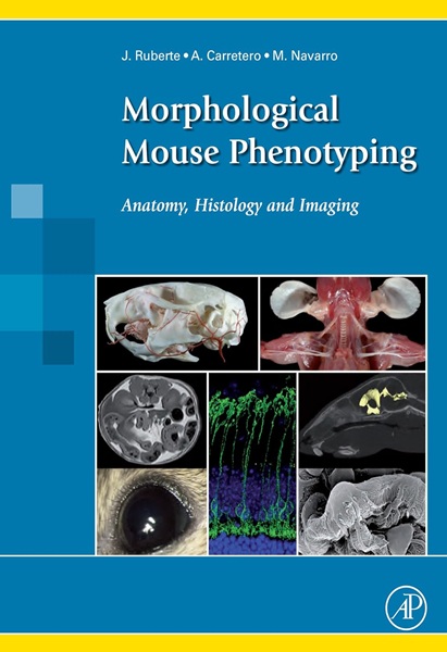
Morphological Mouse Phenotyping: Anatomy, Histology and Imaging is an atlas of explanatory diagrams and text that guides the reader through normal mouse anatomy, histology, and imaging. The book is targeted for mouse researchers and veterinarian and human pathologists, and presents a complete, integrative description of normal mouse morphology. Disease animal models are fundamental in research to improve human health, making the mouse the most used animal disease model. This atlas features more than 2,200 original images showing the anatomy, histology, and cellular structure of mouse organs, and offers an integrative vision of mouse morphology using correlative X-ray, computed tomography, magnetic resonance, and ultrasound images. 585 p.

- Jesus Ruberte. DVM, PhD, Professor, Head of the Mouse Imaging Platform. Center for Animal Biotechnology and Gene Therapy, Department of Animal Health and Anatomy, Veterinary School, Universitat Autonoma de Barcelona (Spain).
- Ana Carretero. DVM, PhD, Associate Professor. Center for Animal Biotechnology and Gene Therapy, Department of Animal Health and Anatomy, Veterinary School, Universitat Autonoma de Barcelona (Spain).
- Marc Navarro. DVM, PhD, Associate Professor. Center for Animal Biotechnology and Gene Therapy, Department of Animal Health and Anatomy, Veterinary School, Universitat Autonoma de Barcelona (Spain).
- Publication date (digital version): 2017-01 – Academic Press.


You must be logged in to submit a review.