Description
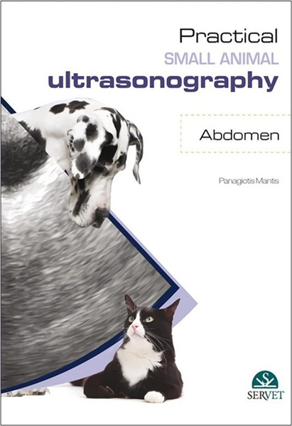
Diagnostic imaging techniques are increasingly important in veterinary medicine. Even if in the past they required a heavy investment, the truth is they are now more affordable and present in almost every vet practice. Radiography was the first technique to arrive and ultrasonography has undoubtedly come to stay. In fact, ultrasonography may provide invaluable information to confirm presumptive diagnosis if, and only if, the clinician is able to perform and identify the normal ultrasonographic appearance of organs, is familiar with variations from normal, and knows how to interpret them. The aim of this ebook Practical Small Animal Ultrasonography: Abdomen, is to provide veterinary surgeons with a visual guide to perform abdominal ultrasonographic examination in dogs, helping them disorders and assisting with the diagnosis and treatment. 262 p.

- Pete Panagiotis Mantis. DVM, European specialist in Veterinary Diagnostic Imaging, Residency in Small Animal Diagnostic Imaging. Currently Senior lecturer in Diagnostic Imaging in the Department of Veterinary Clinical Sciences, Royal Veterinary College, London (UK).
- Publication date (digital version): 2016-04 – Servet (imprint of Grupo Asis); Copyright © 2016 by Grupo Asis Biomedia, S.L.


You must be logged in to submit a review.